
RV-focused acquisition
| View | RV-focused apical four-chamber: from the A4C, rotate slightly counterclockwise at the apex to maximize RV area, keeping the LV apex centred and avoiding anterior tilt into a five-chamber view. |
|---|---|
| Timing | End-diastole for linear dimensions and area; the free wall must be visible throughout the cycle. |
| Caliper / trace | Length from the mid tricuspid annulus to the compacted apex; basal diameter in the basal third; mid diameter at the papillary-muscle level; area traced along the compacted border including trabeculations and moderator band. |
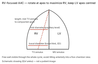
Correct example
Entire right ventricle displayed with the largest basal diameter and the free wall seen through the cycle; measurements made in this view, which is more reproducible than the standard four-chamber view; at least two clips recorded.
Common error
Measuring the RV on the LV-centred four-chamber view, tilting anteriorly into a five-chamber plane, or a plane that misses the free wall; minor transducer rotation changes the RV dimension substantially.
Effect on interpretation
Off-axis views under- or overestimate RV basal diameter and area, so FAC and the dilatation cut-offs are only meaningful on a well-positioned RV-focused view; the view is also the basis for TAPSE alignment and RV strain.
Why
The RV's crescentic shape wrapped around the LV means its apparent size depends on the cut plane; a dedicated, maximized view standardizes the reference points that the normal values were derived from.