
Strain tracking
| View | Three apical views at reduced sector width including only the ventricles, true apex visualized, speckle tracking of the full wall. |
|---|---|
| Timing | Zero strain at mitral valve closure (end-diastole), end-systole at aortic valve closure; in AF average several cycles or use the third of three similar cycles. |
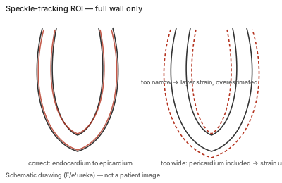
| Caliper / trace | Region of interest covering the full myocardial wall only, drawn straight along the long axis; tracking checked against the underlying motion and loops recorded. |
Correct example
Tracking is inspected segment by segment against the visible myocardial motion; poorly tracked segments are excluded, and if more than three segments are uninterpretable GLS is not reported; serial studies use the same vendor and software.
Common error
Accepting smoothed curves without checking tracking, an ROI that includes pericardium or only a subendocardial layer, foreshortened apical views, or event timing that does not match valve closure.
Effect on interpretation
A wide ROI underestimates strain, a narrow one overestimates it, and foreshortening inflates apical values; because serial change is judged in absolute strain, these errors can mimic or mask a 15% relative deterioration.
Why
GLS is the averaged Lagrangian shortening of the wall across the apical views; speckle tracking regularization can smooth over failed tracking, so visual verification is the only safeguard.