
Aortic jet velocity: CW envelope selection
| View | Continuous-wave Doppler with the dedicated (nonimaging) transducer from multiple windows: apical, right parasternal, suprasternal. |
|---|---|
| Timing | Systolic ejection; average ≥3 beats, ≥5 consecutive beats with irregular rhythm, avoiding post-extrasystolic beats. |
| Caliper / trace | Peak velocity at the outer edge of the dense signal; VTI and mean gradient traced along the outer edge of the dense envelope; report the window that gave the highest velocity. |
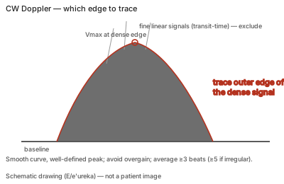
Correct example
A smooth curve with a dense outer edge and a clear peak, recorded with the scale set so the signal fills but fits the display, high wall filter and reduced gain; the highest velocity from any window is reported and lower values from other views are not.
Common error
Including the fine linear transit-time signals above the dense envelope, overgaining, measuring from a single window, applying angle correction, or recording an eccentric MR jet as the AS jet.
Effect on interpretation
Beam misalignment underestimates velocity and, through the squared Bernoulli relationship, underestimates the gradient even more; including fine linear signals or an MR jet overestimates severity.
Why
The curve shape itself carries information: mild obstruction peaks early with a triangular curve, severe stenosis is rounded with a mid-systolic peak, and dynamic subaortic obstruction peaks late.